

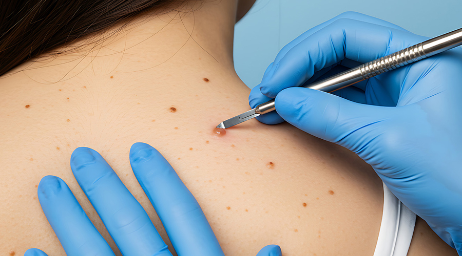
Skin Biopsy (When Indicated)
A skin biopsy is a minor diagnostic procedure in which a small sample of skin tissue is removed and sent for histopathological examination under a microscope. It is an essential tool in dermatology when a clinical diagnosis cannot be confirmed by examination alone, when a skin condition is not responding as expected to treatment, or when a lesion has features that warrant further assessment. A skin biopsy provides definitive diagnostic information that guides appropriate treatment.
When Is a Skin Biopsy Recommended?
A lesion with features suggestive of malignancy or atypical changes that require histopathological confirmation.
Inflammatory skin conditions that are uncertain in diagnosis or not responding to standard treatment.
Suspected scarring alopecia — where scalp biopsy is key to identifying the specific type and guiding treatment urgency.
Unusual or persistent rashes requiring tissue-level diagnosis.
Connective tissue disorders such as lupus or dermatomyositis where skin biopsy supports systemic diagnosis.
Assessment of pigmented lesions that cannot be definitively classified clinically or dermoscopically.
Drug reactions or vasculitis requiring histopathological and immunofluorescence evaluation.
Types of Skin Biopsy
Punch Biopsy
The most commonly performed type. A circular punch tool (typically 3–4 mm) is used to remove a full-thickness core of skin. Provides an excellent sample of the epidermis, dermis, and upper subcutis. The small wound is closed with 1–2 sutures.
Shave Biopsy
A superficial slice of the raised lesion is removed using a scalpel. Suitable for elevated, exophytic, or superficial lesions. Does not provide full-thickness tissue — less suitable for conditions requiring dermal assessment.
Excisional Biopsy
The entire lesion is surgically excised with a margin of normal skin. Both diagnostic and potentially therapeutic — used for small lesions that are completely excised for histopathological examination. Ideal for pigmented lesions where full assessment is needed.
Incisional Biopsy
A wedge or portion of a larger lesion is excised for analysis. Used when the lesion is too large to fully excise or when sampling a representative portion is sufficient for diagnosis.
The Biopsy Procedure
1. Site Selection
The most representative, active, or diagnostically informative area of the lesion is selected for sampling — avoiding necrotic centres, over-treated areas, or sites unlikely to yield diagnostic tissue.
2. Local Anaesthesia & Sampling
Local anaesthetic is injected to ensure a pain-free procedure. The biopsy is performed quickly with minimal discomfort. The sample is placed in appropriate fixative (formalin) and sent to a qualified dermatopathologist for analysis.
3. Wound Care
Punch and excisional biopsies are sutured and sutures removed at 7–10 days. Shave biopsy sites heal by secondary intention with simple wound care. Post-procedure care instructions are provided.
Benefits of Skin Biopsy
Provides definitive histopathological diagnosis guiding precise treatment.
Identifies or rules out malignancy with certainty.
Essential for scarring alopecia diagnosis before irreversible follicular damage occurs.
Guides systemic workup in connective tissue and autoimmune conditions.
Quick, minor procedure performed under local anaesthesia in the clinic.
What to Expect
The biopsy procedure itself takes only a few minutes and is well-tolerated under local anaesthesia. Results from histopathology are typically available within 1–2 weeks. Your dermatologist will review the report with you and discuss diagnosis, implications, and any additional investigations or treatment adjustments required based on the findings. A small scar at the biopsy site is expected and usually fades well over time.